Manufacturer of Optical Spectrometers, Camera Systems for OEM Industrial Applications
OEM gratings design and production capabilities
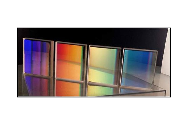
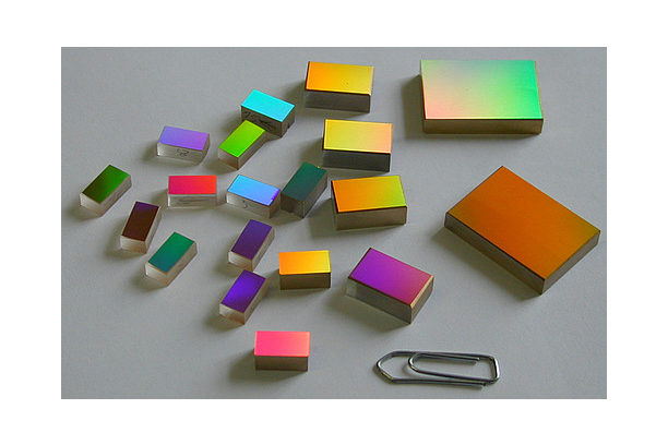
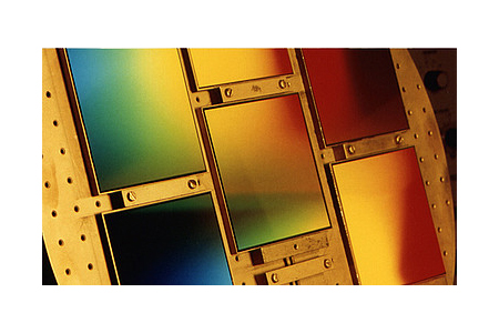
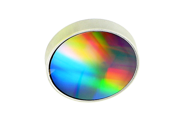
HORIBA Scientific produces a wide range of holographic master gratings from which high precision replicas are manufactured.
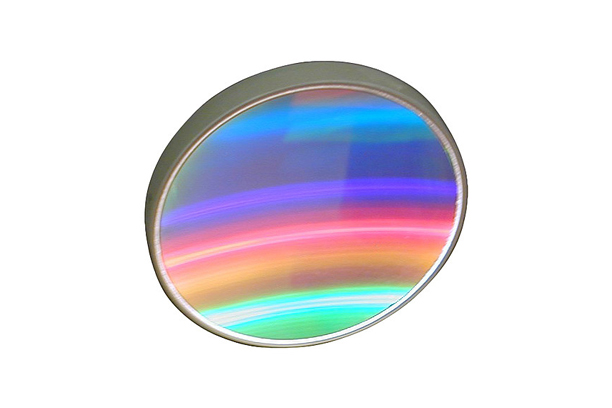
Type IV aberration-corrected flat field and imaging gratings are designed to focus a spectrum onto a plane surface, making them ideal for use with linear or 2-D array detectors.
These gratings are produced with grooves that are neither equis-paced nor parallel, and are computer optimized to form near-perfect images of the entrance slit on the detector plane.
ㆍ Replica gratings from holographic aberration corrected master gratings
ㆍ Holographic Master can be blazed by Ion-beam etching method for higher efficiency
ㆍ Ideal for robust, compact and low stray light spectrometers
ㆍ Several references available (not all are listed in the catalogue)
ㆍ Large range of dispersion available (from few nm/ mm to above 100nm/mm)
ㆍ Multiple spectral range from UV to IR
ㆍ Coating: Al or gold.
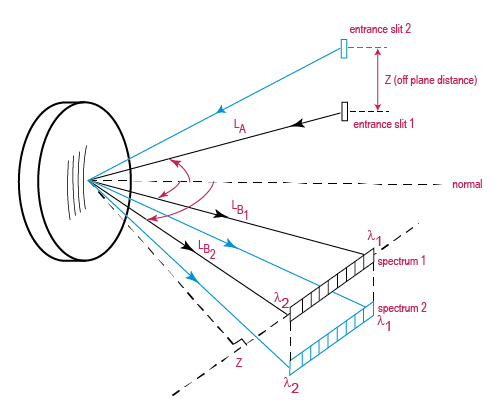
Fig. 1: This illustration shows a “super corrected grating” imaging two independent sources onto two independent linear arrays. Spectrum 1 is a “sample spectrum” from slit 1 and spectrum 2 a reference spectrum from slit 2. These “slits” could be fiber optic inputs
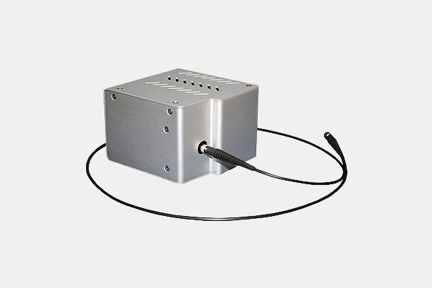
Fig. 2: Typical implementation into a spectrograph
Product Management and Quality
|
Customer Oriented and Confidentiality
|
ㆍ Spectral ranges: from 170 to 4180 nm
ㆍ Average dispersion (nm/mm) : from 1 to 106 nm/mm
ㆍ Diffraction efficiency optimized for UV, or Vis, or NIR
ㆍ Radius of curvature : from 58 to 330 mm
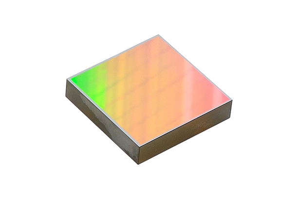
ㆍ Dimensions from 25 x 25 mm
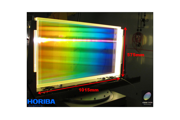
ㆍ For customers in need of larger dimensions, HJY can record a custom-made holographic grating master specifically for replication.
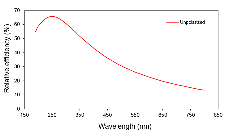
Fig. 3: Typical efficiency curve* for 533 02, 285 l/mm, 190-800 nm
*This efficiency curve is absolute theoretical efficiency, calculated using rigorous electromagnetic theory, taking into account the true groove profiles of manufactured gratings measured with an atomic force microscope (AFM).